This page was created for University of Wisconsin undergraduate course Genetics 564
|
The EFHC1 Protein
EFHC1 protein, or EF Hand Containing 1 protein, was so named because of the EF hand motif found in the structure. An 'EF hand' is a helix-loop-helix structural domain in a protein that allows for the perfect geometry to bind a calcium ion [2]. Figure 1A shows this structure using ribbons to represent amino acid strands, demonstrating the two helices in yellow and blue and the loop connecting them in red. Figure 1B shows why this term is called an EF hand - as the structure closely resembles that of a hand with a calcium binding site right in the middle.
The EFHC1 protein, shown in linear form in the image below, also contains 3 other important domains not included in the namesake, the DM10 domains. Within and between these DM10 regions are where mutations causing JME cluster, as shown in the box below with each line indicating a point mutation. (2) The locations of these DNA mutations are indicated in black arrows in the image. The label for each mutation contains a first letter indicating the wild type amino acid and the final letter indicating the mutated state amino acid that has replaced it. One particular mutation, Argenine 182 to Histidine, is present in 65% of juvenile myoclonic epilepsy cases analyzed and by far the most common mutation. 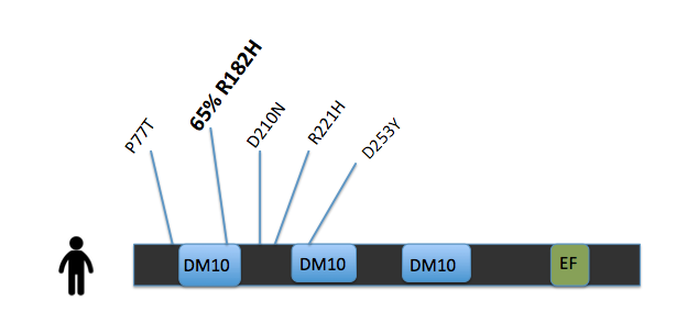
Much is still unknown about the function of EFHC1 within human cells, but the protein has been linked to cell apoptosis (programmed cell death) and mitotic spindle structure. (2) The Ca2+ binding domain has hinted toward involvement of EFHC1 protein in the neuronal firing process. DM10 domain function is yet to be proven through research [3].
|
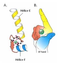
Figure 1. EFHC1 Model of Calcium Binding
|
References
(1) Grisar et al. Myoclonin1/EFHC1 in cell division, neuroblast migration, synapse/dendrite formation in juvenile myoclonic epilepsy. National Center for Biotechnology Information. 2012.
(2) Medina et al. Novel mutations in Myoclonin1/EFHC1 in sporadic and familial juvenile myoclonic epilepsy. Neurology Journal. May 2008. http://www.ncbi.nlm.nih.gov/pubmed/18505993
(3) King, Stephen. Axonemal Protofilament Ribbons, DM10 Domains, and the Link to Juvenile Myoclonic Epilepsy. Cell Motility and the Cytoskeleton. Volume 63. 2006. http://onlinelibrary.wiley.com/store/10.1002/cm.20129/asset/20129_ftp.pdf?v=1&t=hturpcy2&s=ac620ef3194976e78700d12e0d250a790d2c6f20 (3) Image 2. De nils et al. Juvenile myoclonic epilepsy as a possible neurodevelopmental disease: Role of EFHC1 or Myoclonin1. Epilepsy and Behavior. 2013.
